


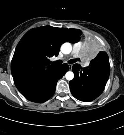
Effusion and collapse CT scan
Hover on/off image to show/hide findings
Tap on/off image to show/hide findings
Effusion and collapse CT scan
- This is a CT slice through the chest of the patient seen in the previous chest X-ray.
- There is narrowing of the left branch bronchi (arrow) with a mass surrounding these (not labelled). The orange area represents the collapsed lungs which has become dense (grey). Compare this with the right lung which still contains air (black). Surrounding the collapsed left lung there is a crescent of fluid - a pleural effusion.
Other anatomical structures
- 1 - Descending thoracic aorta
- 2 - Ascending thoracic aorta (PULLED to left)
- 3 - Main pulmonary artery